Point-of-care ultrasound continues to demonstrate its value in the rapid assessment of patients presenting with acute respiratory symptoms. This recent case highlights how thoracic POCUS can provide immediate diagnostic clarity and guide early management.
Clinical Presentation
A 43-year-old gentleman with a significant smoking history presented with:
- Shortness of breath
- Fever
- Productive cough
- General malaise
The clinical picture initially suggested a lower respiratory tract infection, but further evaluation was required to determine the cause of his respiratory compromise.
POCUS Findings
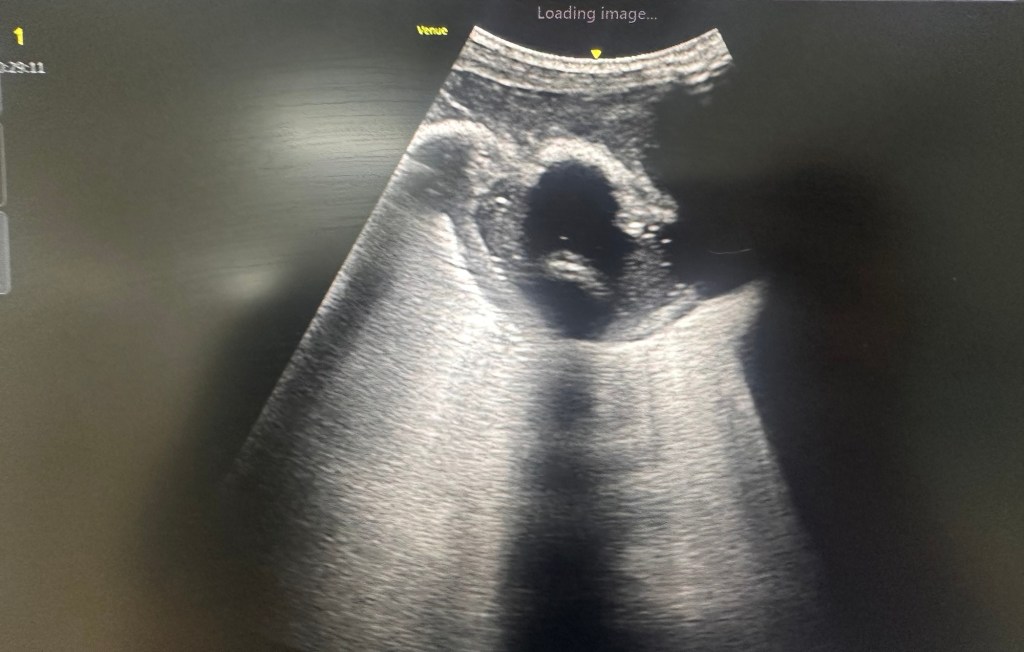
Thoracic ultrasound revealed a complex pleural effusion with internal echogenic debris and loculations. Rather than the simple anechoic appearance seen in uncomplicated pleural effusions, the collection contained multiple internal echoes, raising immediate suspicion for a pleural space infection.
The ultrasound appearance was highly suggestive of empyema.
Ultrasound-Guided Aspiration
A diagnostic thoracentesis was performed under ultrasound guidance.
The aspirated fluid was thick, purulent, and cream-coloured, confirming the suspicion of pleural infection.
The pleural fluid pH could not be analysed by the blood gas machine. This is not uncommon in empyema, where highly viscous and purulent fluid may prevent accurate analysis.
Diagnosis
The combination of:
- Clinical sepsis
- Complex loculated pleural effusion on ultrasound
- Frank pus on aspiration
was diagnostic of empyema.
Management
The patient was commenced on appropriate intravenous antibiotics and referred to the respiratory team. A CT thorax had already been performed to further define the extent of pleural disease and assess for potential intervention.
Given the relatively small but loculated nature of the collection, chest drain insertion may prove technically challenging. Depending on the patient’s clinical progress and multidisciplinary assessment, options may include image-guided drainage, intrapleural therapy, or surgical intervention such as Video-Assisted Thoracoscopic Surgery (VATS).
Learning Points
Thoracic POCUS rapidly differentiates simple from complex pleural effusions.
Internal echoes, septations and loculations should raise suspicion for empyema.
Frank pus obtained during thoracentesis is diagnostic of empyema regardless of pleural fluid pH.
Ultrasound-guided procedures improve diagnostic accuracy and procedural safety.
Early identification allows prompt antibiotic therapy and specialist referral.
Why This Case Matters
This case is another example of how POCUS brings imaging directly to the bedside, allowing clinicians to move quickly from diagnostic uncertainty to targeted management. What initially appeared to be a routine respiratory presentation was rapidly identified as a pleural space infection requiring urgent treatment.
POCUS doesn’t replace clinical assessment—it enhances it, often providing the missing piece of the puzzle within minutes.
Images and case details shared with appropriate consent and anonymisation.

Leave a comment